VCT 2D Materials Lab at UTokyo (400 m2) features state-of-art equipment for epitaxy synthesis (12 CVD furnaces), and characterizations of 2D layered materials. 


Ti-Premier-Tool-Image-v3.jpg

Brand: Bruker

Model: TI Premier

Bruker's Hysitron TI Premier Series was specifically designed to deliver industry-leading, quantitative nanomechanical characterization within a compact platform. Built upon proven Hysitron technology, the Hysitron TI Premier provides an essential toolkit for your nanoscale mechanical and tribological testing. Routine measurements to advance research can be accomplished utilizing the versatile base configurations of the Hysitron TI Premier, while numerous technique upgrade options are available to meet the potential diversity of your future characterization needs.


Screen Shot 2018-05-19 at 10.07.10 AM.png

Brand: KSV NIMA

Model: MicroBAM

MicroBAM enables the visualization of Langmuir monolayers or adsorbate films at the air-water interface for example as a function of packing density. 

  • Monolayer/film homogeneity. When combined with a KSV NIMA L & LB Trough, observation can be performed during compression/expansion at known surface pressures. Verifying the homogeneity of the monolayer already prior to deposition will speed up the research considerably.

  • Optimizing the deposition parameters. For a high quality LB coating, the measurement parameters such as compression speed, waiting times, temperature and subphase content need to be optimized. With MicroBAM it is easy to study the effects of these parameters and choose the optimal conditions.

Langmuir layer structure and behavior studies

  • Monolayer/film behavior. It is possible to observe phase changes, phase separation, domain size, shape and packing.

  • Monitoring of surface reactions. For example, photochemical reactions, polymerization reactions as well as enzyme kinetics can be followed in real-time.

  • Monitoring and detection of surface active materials. For example protein adsorption and nanoparticle flotation.


Confocal Raman from WITec

alpha300R-motorisiert-095-neu-bearbeitet.png

Brand: WITec
Model: Alpha300AR+

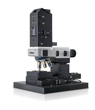
The WITec Raman microscope and imaging system combines an extremely sensitive confocal microscope with an ultrahigh-throughput spectroscopy system for unprecedented chemical sensitivity. A highly efficient combination of optical components such as filters, objectives and lenses used in conjunction with the most sensitive detectors available provide the highest spatial and spectral resolution.

The Raman system has a high throughput configuration, with the following specifications and modes of operation:

    • Raman Spectral Imaging: acquisition of complete Raman spectra at every pixel (mapping)
    • Acquisition of Raman spectra at selected areas (micro-Raman)
    • 3D confocal Raman imaging
    • Confocal microscopy in reflection
    • Confocal fluorescence imaging
    • Optical resolution: diffraction limited lateral typ. 250 nm @ 532 nm excitation wavelength (with objective NA 1.4)
    • Sample size: max. 120 mm in x- and y-direction, 25 mm in height (for samples with larger height an adapter can be used)
    • Wavelength range VIS and NIR, others optional, typ. detection from < 95 -ca. 4000 wavenumbers @ 532 nm excitation wavelength
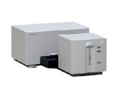
    • Optimized spectrometer and CCD detector each for VIS and NIR

 Research grade optical microscope with 6x turret:

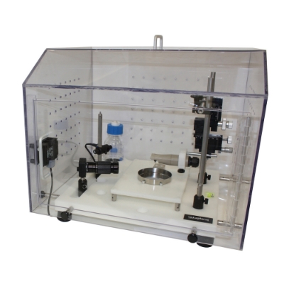
     • Piezo-driven scan platform for sample scanning, continuous scan range up to 200um in x- and y-direction, 20um in z-direction, scan    
        hardware linearized with closed-loop feedback, scan resolution: position accuracy < 2nm in x- and y-direction and < 0.2nm in z- direction, 
        linearity better than 0.03%, achieved through capacitively controlled hardware linearization
     • Video system: eyepiece color video camera
     • Motorized z-stage system for automated approach, 30mm travel, single step 10 nm resolution
     • Manual sample positioning in x- and y-direction, 20mm travel, gradation10um, resolution < 1um
     • LED white-light source for Köhler illumination
     • AFM extension with contact, lateral force and AC modes


banner-20th-anniversary-2145.jpg

Brand: PARK XE-100 AFM

Model: XE-100 AFM with optical head and Hysitron Triboscope adapter head

The XE-100 AFM provides the ultimate AFM/SPM solution for Non-Contact nanoscale metrology of small samples in data storage, semiconductors, materials science, polymers, electro chemistry and other applications in nanoscience and engineering.

XE-100 AFM with optical head that is capable of:

     •  True Non-Contact Mode

     •  Contact mode

     •  Intermittent mode

     •  Lateral Force Microscopy

     •  Force – distance spectroscopy


FullSizeRender.jpg

Brand: Signatone/Lakeshore Integrated Probe Station

Model: 100

For parametric testing that maximizes the productivity of technicians and engineers in R&D, the Model 4200-SCS Semiconductor Characterization System combines lab-grade DC and pulse device characterization, real-time plotting, and analysis with high precision and sub-femto amp resolution in a fully integrated characterization system. Options include the Model 4210-CVU Capacitance-Voltage Unit, Model 4225-PMU Ultra-Fast I-V Module, Model 4220-PGU Pulse Generator Unit, and Model 4225-RPM Remote Amplifier/Switch.


QE-R IPCE  System

From Enli Tech. Inc (Taiwan), is a multifunctional work station for Photovoltaic measurement in tandem with determining the absolute external quantum efficiency (E.Q.E.) of photovoltaic device. It’s now used by target  research center and solar  cell   manufacturers  to improve the  conversion  efficiency.  


Brand: Agilent

Model: UV/Vis Spectroscopy 8453

The Agilent 8453 UV-Visible spectrophotometer uses a photodiode array (PDA) for simultaneous measurement of the complete ultra-violet to visible light spectrum in less than one second. The PDA technique brings exceptional reliability and repeatability.


Spraybase High Throughput Electromechanical 3D Assembly

Electromechanical Assembly enables the synergistic coupling of dissimilar functional building blocks to afford unprecedented material properties at nano-to-micro scale. 


Leica DM-2500 Optical Microscopy

The Leica DM2500 microscope is the ultimate tool for demanding tasks in pathology, cytology, haematology, and even for basic research applications. With its powerful 100W illumination, high-quality optical performance,  and state of the art accessories, the Leica DM2500 is especially well suited for nanotechnology that requires differential interference contrast or high-performance fluorescence.